Spine Model

Validated small and large animal models are available to study spinal fusion at the cervical, thoracic and lumbar levels using anterior or posterior approaches.
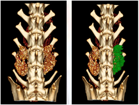
CT scan view of dog PLF model 4 weeks post-surgery
(green: bone volume calculation)

Species:  
  1. ● Rabbit
  2. ● Rat
  3. ● Dog
  4. ● Goat and sheep
  5. ● Pig
  6. ● Primate

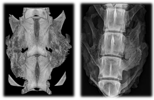
View of dog PLF model 12 weeks post-surgery (left: Micro CT, right: Faxitron)

Types of Models

Procedures are usually performed on one level but multilevel fusions are also possible:

  1. ● Anterior Cervical Fusion
  2. ● Anterior Cervical Discectomy (sheep, goat)
  3. ● Posterolateral Lumbar Fusion PLF (rat, rabbit, dog, sheep, pig, primate)
  4. ● Posterior Thoracic Fusion (rat)
  5. ● Posterolateral Interbody Fusion (dog, sheep)

Outcome Measurements:

  1. ● Radiography: Intervertebral Angle
  2. ● CT and Micro CT (fusion status and bone volume)
  3. ● High-Resolution X-Rays (Faxitron)
  4. ● Non-Invasive Biomechanical Testing
  5. ● DEXA: Bone Mineral Density
  6. ● Histology and Histomorphometry: Bone Quality

PMMA histology on a dog PLF model (top) and a rabbit PLF model (bottom).
0 +
Customers Around
The World
0 +
Countries Served
0 +
Due Diligence
0
Preclinical Histology Slides Performed

Get In Touch With Us

Thank You!!

We will get back to you shortly.