Despite these limitations, small and large animal models have been developed to study the effects of bone substitutes, scaffold, biological products or cell-based products on bone fracture repair.
Fracture bone healing is usually an optimal biological process. However, delayed healing or non-union can occur on patients for multiple reasons. In addition, bone fractures are more frequent and more problematic in osteoporotic patients as the bone structure is compromised. Fracture model, segmental and critical-size defect models have been developed in several species.
Animal Models
Critical-size defects:
- ● Calvarias critical-size defects in rat and rabbit
- ● Mandibular critical-size defects in mini-pig and dog
- ● Femur critical-size defects in rat
- ● Ulna critical-size defects in rabbit
- ● Tibial critical-size defects in sheep and mini-pig
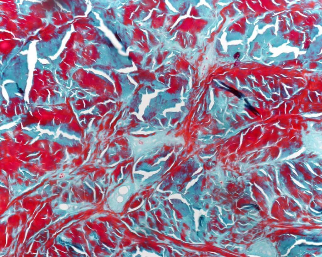
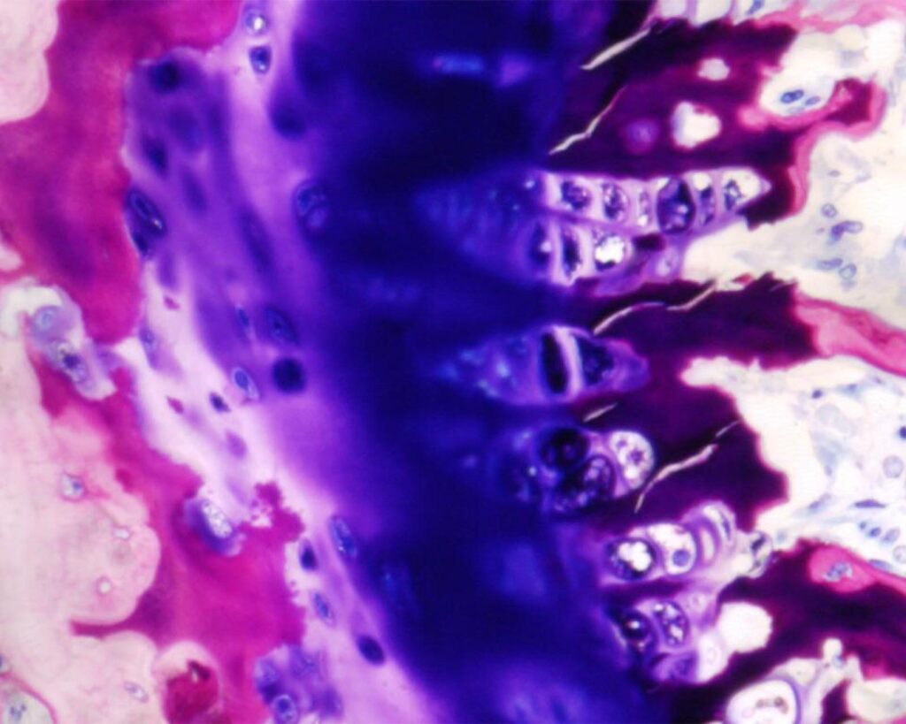
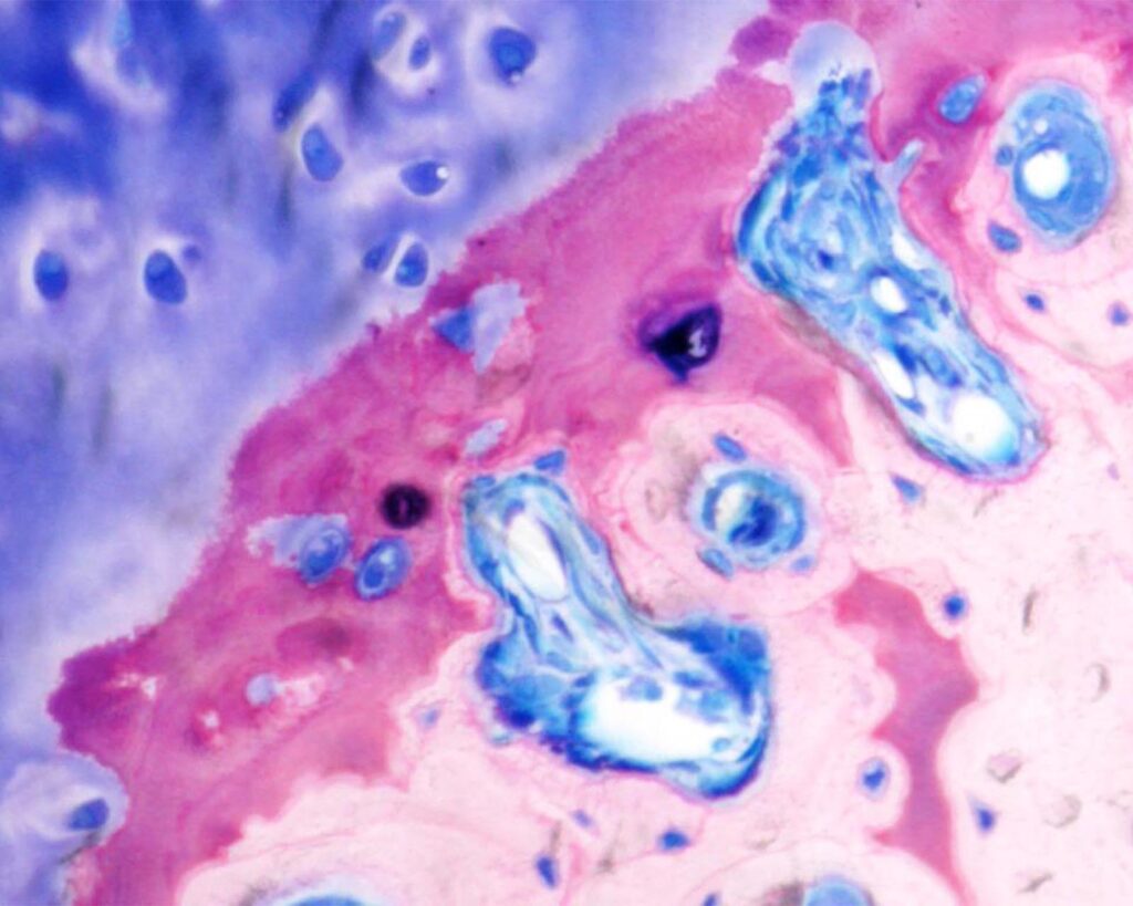
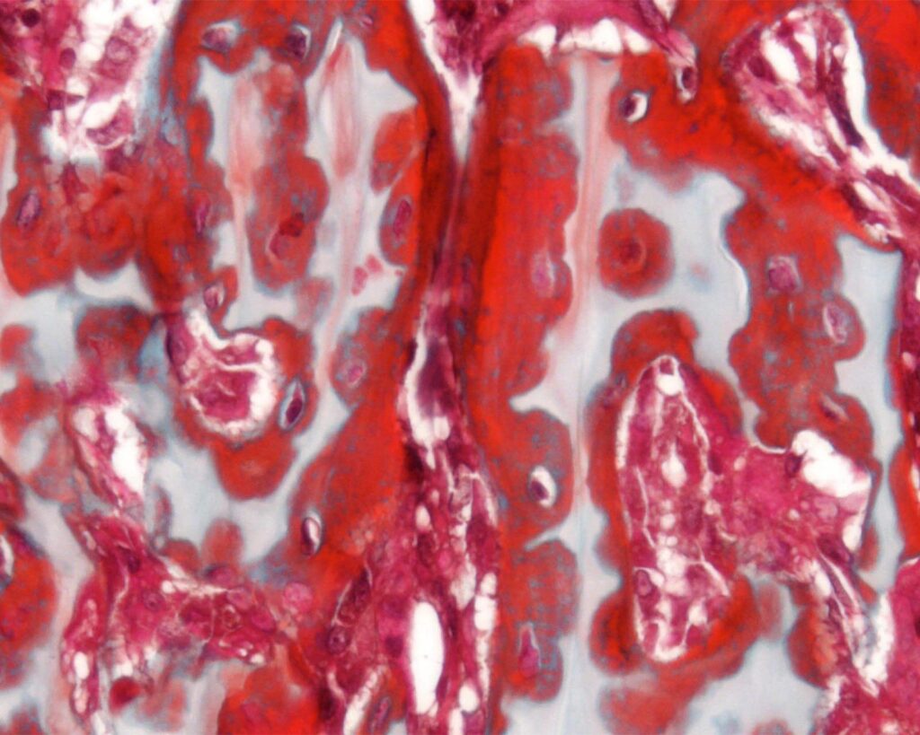
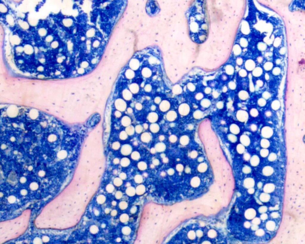
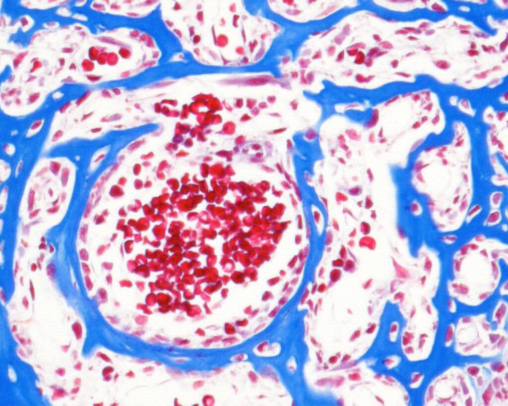
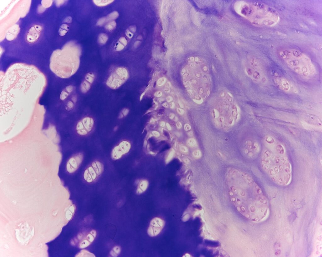
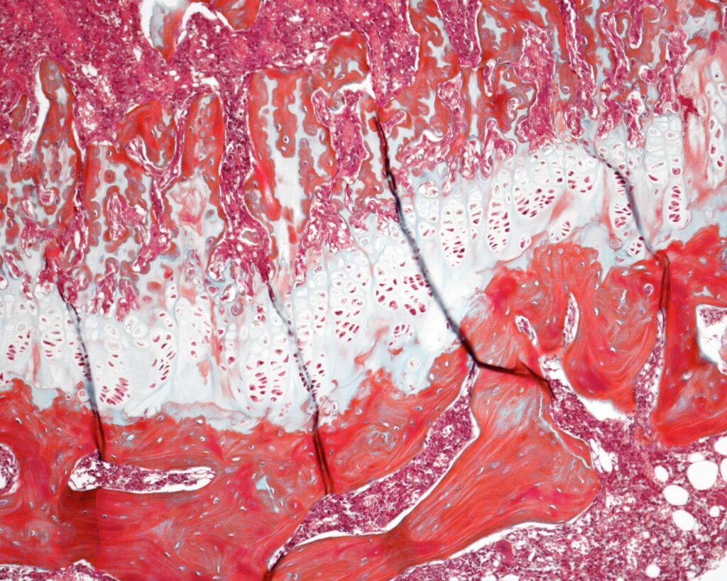
Outcome Measurements:
- ● In vivo and ex vivo BMD and BMC measurements of cortical and trabecular bones by DEXA, pQCT and microCT
- ● Biomechanical testing
- ● Physiological bone turnover markers (blood, urine)
- ● Ash chemical analysis
- ● Histomorphometry, histology